📘 ❞ ATLAS OF HUMAN SKELETAL ❝ كتاب
كمال الاجسام - 📖 ❞ كتاب ATLAS OF HUMAN SKELETAL ❝ 📖
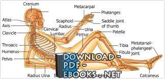
█ _ 0 حصريا كتاب ATLAS OF HUMAN SKELETAL 2024 SKELETAL: this image shows the skeleton of our body (the bones that forms and supports body) showing: 1 cranium 2 atlas vertebra 3 axis vertebra 4 clavicle 5 thoracic vertebra 6 humerus 7 pelvic bone 8 lumbar vertebra 9 cervical vertebra 10 radius bone 11 ulna bone 12 scaphoid bone 13 metacarpal bones 14 phalanges fingers 15 saddle joint thumb 16 sacral vertebrae 17 coccys vertebra 18 head femur 19 femur bone 20 patella 21 tibia bone 22 fibula bone 23 metatarsal phalangeal joint 24 calcaneus bone Illustration provided by: Rolin Graphics In chapter we will introduce skull their topographic relations to each other With exception of Mandibula lower jaw), bone is connected by tight immovable joints neighborhood The areas contact between two are called sutures ( Suturae ), which represent thin junctions irregular interlocking edges adjacent adults Suturae consist connective tissue We use many different views in show all structures To understand it important know, position or orientation defined as standard set up a nomenclature for orientations skulls anatomy horizontal plane was Frankfort more than hundred years ago The Horizontal plane tree points: bottom margin left eye cavity top margins external auditory porus (see 2 6 Os temporale) on both sides Fig 4 : anatomic perspectives parallel perpendicular (Frankfort Horizontal) Perspective Explanation Norma frontalis View at frontal outer surface occipitalis back (posterior) lateralis right from side verticalis basalis inferior 5 Standard describe structures, also make some cuts through inner parts visible additional most focus major Following the Suturae borders using knowledge chapter’s topography should be easy you learn For detailed descriptions see following chapters deal with separate FH ANATOMY J ARTNER ET AL 2002, WWW JURAJARTNER COM 5PAGE Front view ( frontalis ) facial ( Viscerocranium ) ( Os frontale ), already belongs the Neurocranium , top: prominent frontale ) top, zygomatic arches laterally, mandible jaw, Mandibula ) bottom, orbital excavations ( Orbitae ) below bone, anterior nasal aperture Apertura nasalis , piriformis ) located middle line cavities, dentition, upper Maxilla ) jaw bone ( Os articulates downwards (on sides) ( Ossa nasalia, sing nasale ), lateral with Maxillae , lacrimal lacrimalia, Os lacrimale ) zygomatica, zygomaticum ) frontale and Ossa nasalia joined one another almost frontonasal suture ( Sutura frontonasalis ) frontomaxillar frontomaxillaris ) unites (Maxilla) connection (suture) arched of Os have to Os zygomaticum Both form together Maxilla exterior (orbital or Orbita ) 7 Orbita) avoid an information overload later illustrations At moment only note, above mentioned extend into interior, where they complex connections four (Os lacrimale, ethmoidale, sphenoidale, palatinum) pyramidal cavities frontale (1), nasale (2), Maxilla (3), zygomaticum (4), Mandibula (5), Orbita (6), ant (7) bony bridge nose united vertical internasal internasalis ) midline anterior or approximately cranium, nasalia ) both Maxillae Divided septum entrance shells ( Conchae ) ( Septum nasi ) within row dentition (teeth) separated by Sutura intermaxillaris under Mandibula ), movable (if small ossicles ear not counted) 1 3 7 6PAGE Fig Left (2) (3) margins, Sutura frontomaxillaris (a), frontozygomatica (b) zygo maticomaxillaris (c) them Parts sphenoidale lacrimale palatinum ethmoidale (5) described Orbita, frontoethmoidalis (e), sphenofrontalis (d), sphenozygomatica (f), (c), ethmoido maxillaris, frontolacrimalis, palato maxillaris palatoethmoidalis Two large apertures can seen cavity: Fissura orbitalis superior (Fs) connects internal cranial cavity, passed Nervus ophthalmicus, occulomotorius, trochlearis, abducens Vena ophthalmica (Fi), part zygomaticus, infraorbitalis corresponding vessels كمال الاجسام مجاناً PDF اونلاين الأجسام (بالإنجليزية: Bodybuilding)؛ لعبة رياضية نشأت أواخر القرن التاسع عشر الميلادي وبدايات العشرين وشرط اساسى فى قانون يكون هناك تناسق عضلى للاعب مبدؤها تضخيم عضلات الجسم وإبرازها واستعراضها مقارنة بعضلات المنافسين الآخرين كتب تتحدث عن رياضة مقدمة الأجسام منذ القدم كان التمجيد من نصيب المكتملة القوية مفتولة العضلات قال ويل ديورانت واصفاً جلجامش الأسطورة الرافدية: «هو طويل القامة ضخم مفتول جريء مقدام جميل يفتن الناس بجماله لا يماثله أحد صورة جسمه يرى جميع الأشياء ولو كانت أطراف العالم كل شيء وعرف واطلع الأسرار واخترق ستار الحكمة الذي يحجب ورأى ما خافياً وكشف الغطاء عما مغطى وجاء بأخبار الأيام التي قبل الطوفان وسار طريق بعيد كابد فيه المشاق والآلام ثم كتب لوح حجري قام به الأعمال» في مصر القديمة الملوك والآلهة ينحتون بالتماثيل تجسدهم مفتولي ممشوقي القوام مشدودي دليلاً القوة والكمال والجمال ينبغي أن عليه الإنسان لدى اليونان البطل الخارق هرقل نصف الإله ونصف البشر يتمتع بجسم خارق للعادة قوي البنيان وقد أصبح هذا بقوته وعضلاته رمزاً للألعاب اليونانية جبل الأولمب قرب أثينا وتبعهم ذلك الرومانيون اقتفوا آثار اليونانيين خطوة بخطوة مع تغيير الأسماء في الهند والصين كذلك النحاتون يتفننون بنحت البشرية بكامل رونقها وقوتها ويصورون أساطيرهم نماذج الكمال الجسماني والعضلي والعقلي لأبطالهم وآلهتهم حد سواء الكتاب المقدس وردت إشارات إلى قوة الأنبياء والقادة لعل أبرزهم الشعبي شمشون الجبار بقوة جبارة وجسد الحضارة العربية الإسلامية أشار القرآن ميزة الكامل القوي بقوله طالوت زعيم بني إسرائيل وقته: (قال إن الله اصطفاه عليكم وزاده بسطة العلم والجسم والله يؤتي ملكه يشاء واسع عليم) البقرة 247 وكذلك قوله موسى النبي: (قالت إحداهما يا أبت استأجره خير استأجرت الأمين) القصص 26 كذلك حفلت الكتب والإسلامية بوصف النبي والصحابة والخلفاء والسلاطين والحكام؛ ووصف أجسادهم ومدى قوتها وصلابتها ومقارنة الواحد منهم بالعشرات الرجال العاديين وعلى الرغم قلة التصاوير والتماثيل المنتسبة إلا قصص ألف ليلة وليلة (الأساطير المثالية) ضجت بالأبطال الخارقين بأجسامهم وقواهم الخارقة سيما السندباد وعلاء الدين وفي عصر النهضة الأوروبية تابع الفنانون أسلافهم والرومانيين بتمجيد الأجساد الممشوقة الكاملة والقوة فصوروا الآلهة والأنبياء والقديسين بأجسام ممشوقة بارزة الرجولة الشعبية أجمع يمجد العضلية والنشاط والصحة البدنية فعندنا الأبطال الشعبيون ثور والبرابرة الجبارون وزينا
this image shows the skeleton of our body (the bones that forms and supports our body)
showing:
1. cranium
2. atlas vertebra
3. axis vertebra
4. clavicle
5. thoracic vertebra
6. humerus
7. pelvic bone
8. lumbar vertebra
9. cervical vertebra
10. radius bone
11. ulna bone
12. scaphoid bone
13. metacarpal bones
14. phalanges of the fingers
15. saddle joint of the thumb
16. sacral vertebrae
17. coccys vertebra
18. head of femur
19. femur bone
20. patella
21. tibia bone
22. fibula bone
23. metatarsal-phalangeal joint
24. calcaneus bone
Illustration provided by: Rolin Graphics
In this chapter we will introduce the bones of skull and their topographic relations to each other. With the exception of
Mandibula
(the lower jaw), each skull- bone is connected by tight immovable joints to the bones of the neighborhood. The areas of contact between two bones are called sutures (
Suturae
), which represent thin junctions between the irregular interlocking edges of adjacent skull bones. In adults
Suturae
consist of tight connective tissue. We will use many different views in this chapter to show all skull structures. To understand the views it is important to know, which position or orientation is defined as the standard. To set up a standard nomenclature for orientations of skulls in anatomy a horizontal plane was defined in Frankfort more than hundred years ago. The
Frankfort Horizontal plane
is defined by tree points: the bottom margin of the left eye cavity and the top margins of the external auditory porus (see chapter 2.6. Os temporale) on both sides. Fig. 2.4.: Frankfort horizontal plane The standard anatomic perspectives are parallel or perpendicular to this (Frankfort Horizontal) plane. Perspective Explanation Norma frontalis View at the frontal outer surface of skull Norma occipitalis View at the back (posterior) outer surface of skull Norma lateralis View at the left or right outer surface of skull from side Norma verticalis View at the top outer surface of skull Norma basalis View at the inferior outer surface of skull Fig. 2.5.: Standard perspectives To describe all skull structures, it is also important to make some cuts through the skull and make the inner parts visible. In this chapter we will use the standard perspectives and additional views to show most skull structures. We will focus on major and important structures to show their topographic relations to each other. Following the
Suturae
as borders and using the knowledge of this chapter’s topography it should be easy for you to learn and to describe all bones of skull. For more detailed descriptions see the following chapters which deal with separate skull bones.
FH
ATLAS OF HUMAN SKELETAL ANATOMY
J.ARTNER ET AL. 2002, WWW.JURAJARTNER.COM 5PAGE
Front view (
Norma frontalis
) of the skull shows the bones of the facial skull (
Viscerocranium
) and the frontal bone (
Os frontale
), which already belongs to the
Neurocranium
, at the top: The most prominent structures in this view are the frontal bone (
Os frontale
) at the top, the zygomatic arches laterally, the mandible (the lower jaw,
Mandibula
) at the bottom, both orbital excavations (
Orbitae
) below the frontal bone, the anterior nasal aperture (
Apertura nasalis anterior
,
Apertura piriformis
) located in the middle line between both orbital cavities, and the dentition, which belongs to the upper (
Maxilla
) and the lower jaw (
Mandibula
). The frontal bone
(
Os frontale
) articulates downwards (on both sides) with the nasal bones (
Ossa nasalia, sing. Os nasale
), more lateral with
Maxillae
, the lacrimal bones (
Ossa lacrimalia, sing.
Os lacrimale
) and with the zygomatic bones (
Ossa zygomatica, sing.
Os zygomaticum
).
Os frontale
and
Ossa nasalia
are joined to one another by the almost horizontal frontonasal suture (
Sutura frontonasalis
). The frontomaxillar suture (
Sutura frontomaxillaris
) unites the upper jaw (Maxilla) with the frontal bone on both sides of the frontonasal connection (suture). The lateral arched parts of
Os frontale
have contact to
Os zygomaticum
. Both bones form together with the upper parts of
Maxilla
the exterior margins of the eye cavity (orbital cavity or
Orbita
) on both sides (see Fig.2.7.: Orbita). To avoid an information overload we will describe the orbital structures later in separate illustrations. At this moment it is only important to note, that the above mentioned bones extend into the orbital interior, where they form in complex connections with four other bones (Os lacrimale, Os ethmoidale, Os sphenoidale, Os palatinum) the pyramidal orbital cavities. Fig. 2.6.: Norma frontalis of skull Os frontale (1), Os nasale (2), Maxilla (3), Os zygomaticum (4), Mandibula (5), Orbita (6), Apertura nasalis ant. (7) The two nasal bones which form the bony bridge of the nose are united by the vertical internasal suture (
Sutura internasalis
) in the midline. The anterior nasal aperture (
Apertura nasalis anterior
or
Apertura piriformis
) is located approximately in the middle of the facial cranium, below both nasal bones (
Ossa nasalia
) and between both
Maxillae
. Divided in the middle by the nasal septum it forms the entrance into the bony nasal cavity with visible nasal shells (
Conchae
) and nasal septum (
Septum nasi
) within. The upper row of dentition (teeth) belongs to both
Maxillae
, which are separated by
Sutura intermaxillaris
under the nasal aperture. The lower row of dentition belongs to the lower jaw (
Mandibula
), which is the only movable bone of skull (if the small auditory ossicles of the middle- ear are not counted).
1 2 3 4 5 6 7
ATLAS OF HUMAN SKELETAL ANATOMY
J.ARTNER ET AL. 2002, WWW.JURAJARTNER.COM 6PAGE
Fig. 2.7.: Left Orbita Os frontale (1), Maxilla (2) and Os zygomaticum (3) form the exterior margins, with Sutura frontomaxillaris (a), frontozygomatica (b) and zygo-maticomaxillaris (c) between them. Parts of Os sphenoidale (4), Os lacrimale (6), Os palatinum (7) and Os ethmoidale (5) form together with the already described bones the bottom of Orbita, separated by Sutura frontoethmoidalis (e), sphenofrontalis (d), sphenozygomatica (f), zygo-maticomaxillaris (c), ethmoido-maxillaris, frontolacrimalis, palato-maxillaris and palatoethmoidalis. Two large apertures can be seen at the bottom of the orbital cavity: Fissura orbitalis superior (Fs) connects the orbital cavity with the internal cranial cavity, passed by Nervus ophthalmicus, occulomotorius, trochlearis, Nervus abducens and Vena ophthalmica superior. Fissura orbitalis inferior (Fi), located between Os sphenoidale and the upper orbital part of Maxilla is passed by Nervus zygomaticus, Nervus infraorbitalis and their corresponding vessels
#2K
1 مشاهدة هذا اليوم#15K
36 مشاهدة هذا الشهر#3K
47K إجمالي المشاهدات-
🎁 كن أول كاتب اقتباس في هذه الصفحة واحصل على هديّة 15 من النقاط فوراً 🎁

